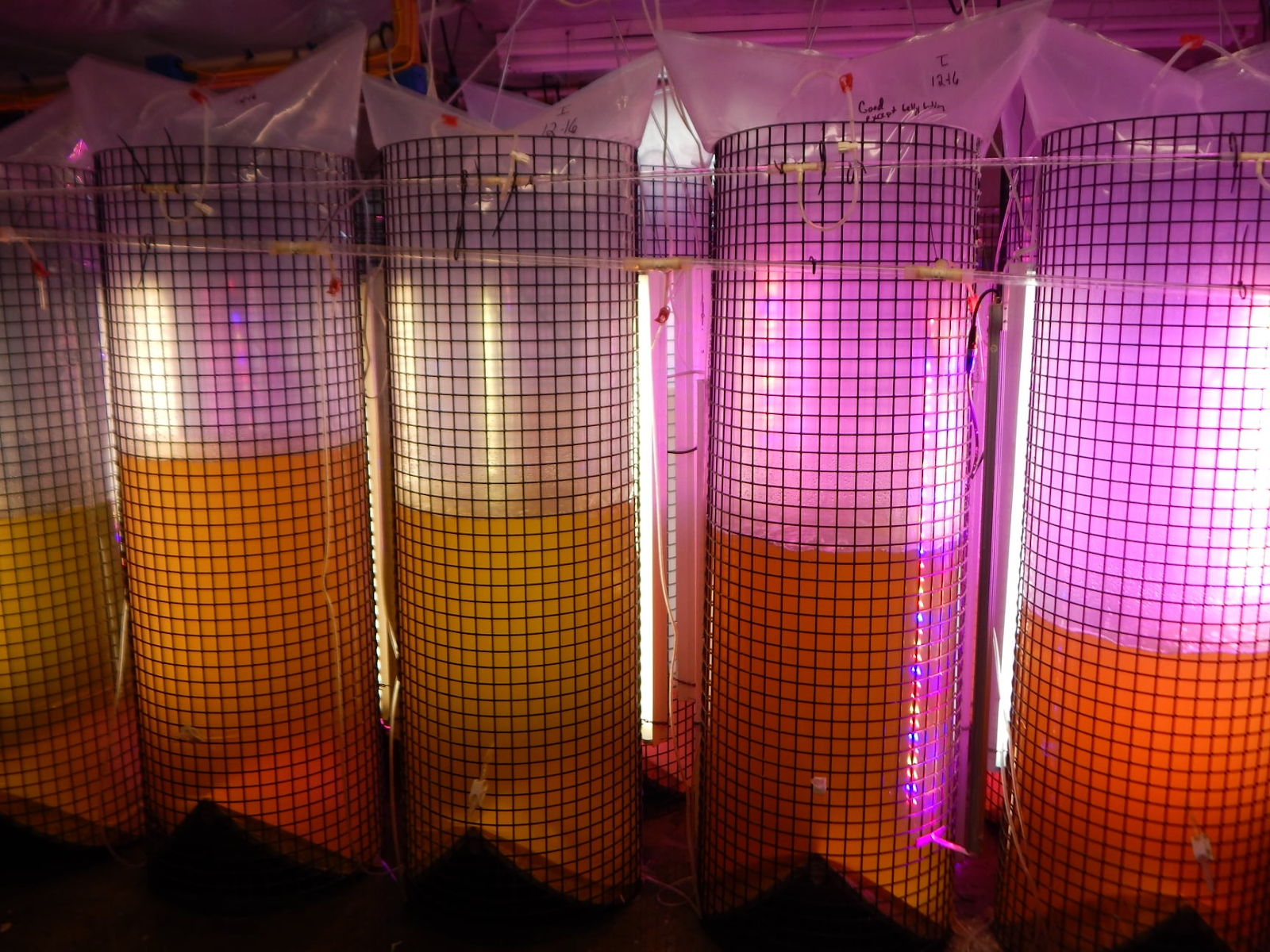
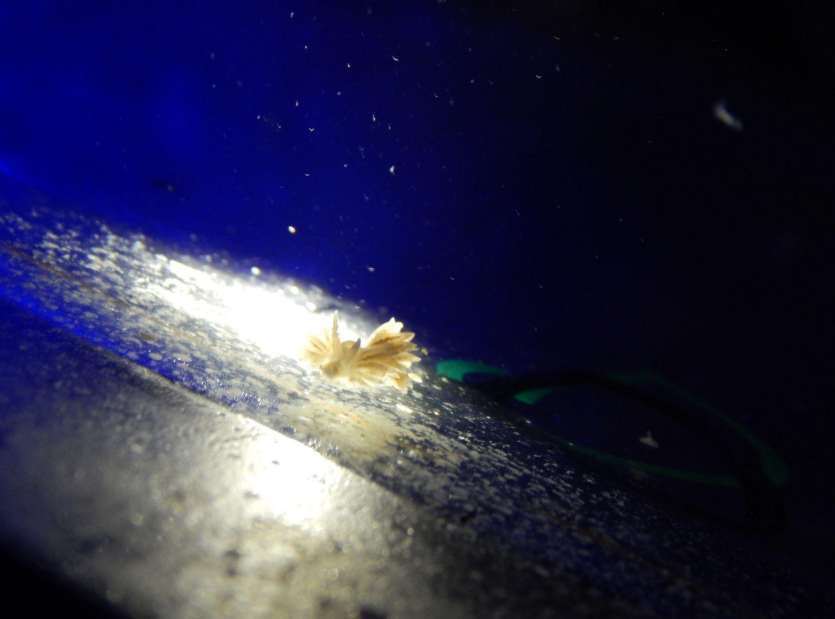
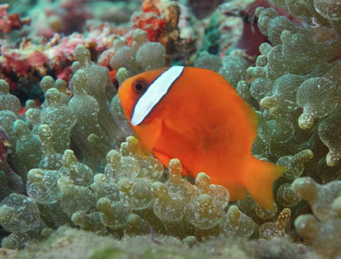
White Corals, Part III: Coral
Disease "White Syndromes"
Proud sponsor of this column
In the first article in
this series, I discussed the phenomenon of coral bleaching and how it can result in a pale
or white-appearing coral. I also compared it to actual tissue loss in corals, showing how
the two can resemble each other. In the second part, I covered a number of events,
organisms, and conditions that could resemble both bleaching and coral disease, resulting
in tissue loss or producing white areas on corals that are difficult to distinguish as to
their cause. In this last article, I will treat the subject of the coral diseases known
collectively as "White Syndromes" (Antonius 1995).
Coral diseases can be quite different in the
gross appearance of the diseased organism. In some cases, the signs are fairly distinct,
while in others they are so similar that there is no way to tell them apart. This becomes
even more problematic given that the etiology or set of conditions that cause and factor
into the disease condition, may not be well established or known at all. In numerous
diseases, the pathological condition that is observed consists of an area of bleached
tissue or an area of recent tissue loss so that the normally pigmented tissue presents a
sharp contrast to the white recently exposed skeleton.
The area that borders the
healthy tissue, including any degenerating tissue and, usually, the area of white exposed
skeleton, is called the "band line." The band line may be quite broad, or very
narrow. It may make sense that a coral disease with a slow progression will generally have
a narrow band line, since other flora and fauna, obscuring the white color soon overtake
the white exposed skeleton. In contrast, a rapidly progressing disease typically has a
wide band line as the tissue loss progresses quickly, leaving more skeleton exposed before
flora and fauna can overgrow the exposed white skeleton.
In some cases, disease may result in
bleaching, either locally or across areas of the colony. Conversely, bleaching can
increase susceptibility to disease. In some diseases (for example, Yellow Blotch Disease),
the band line may have bleached tissue and areas of exposed skeleton. This particular
disease tends to move very slowly, and yet has a wide disease band. This is because of the
bleached regions that can extend upwards of several inches into the tissue that still
covers the skeleton. Thus, each instance of disease must be carefully considered and
observed over time.
The band line is important in another way.
The rate of progression of white syndromes may be partially diagnostic in distinguishing
the type of disease present. For example, White Band Disease (WBD) tends to move fairly
slowly across the coral surface, whereas White Plague Type II (WPII) tends to move quite
rapidly. Therefore, measuring the rate of tissue loss and the width of the band line can
help a potential investigator to determine which of these two grossly similar diseases may
be at work.
Becoming Familiar with the White
Syndromes:
There has been an increase in the reports of
coral diseases over the past twenty years. The first white syndrome identified was WBD
(Gladfelter 1977). This was followed by White Plague in the same year (Dustan 1977). Since
then, a host of other white pathologies have been reported. For many of these, the cause
or causative agent has yet to be identified, or has been tentatively identified. It is not
yet known for certain if some of these diseases are the same, or variations of, another
condition. It is equally possible that different and as yet unnamed diseases are being
lumped into categories of currently named diseases because of a lack of proper study. For
most coral diseases, the state of knowledge is greatly lacking. This may be even truer of
the white syndromes. A table of currently named diseases with signs of white or somewhat
white (pale) areas, and summarizing information about them, is shown below (Table 1). A
photo gallery of some of the white syndromes is provided at the end of the article
(Appendix 1).
Proud sponsor of this column
Table 1:
Name
Causative Agent(s)
Reference
Description
White
Band Disease Type I (WBDI)
Unknown
possible role of gram negative ovoid bacterial bodies in tissues of some samples
(Peters et al. 1983)
In some of the diseases, one or more
white "lines" indicating a band line may occur across a coral surface or around
a colony or branch. Others appear as spots or patches on the coral surface, with the band
line moving outward from a central focus or initiation point. In others, the result is
bleaching or even colony-wide tissue release.
Factors in White Syndromes:
Because so little is known about the cause
and pathology of the many diseases, it is difficult to determine what the primary or
secondary causes might be, including the influences of various stressors. In particular,
there is a great deal of question as to the roles of pathogens and other biotic factors
versus environmental stressors (abiotic factors). There is probably significant
interaction between numerous factors to produce a disease condition. In seems to be a
general consensus that for most coral disease, new pathogens never before seen are not to
blame. Rather, the microbial ecology of corals is coming under study, and research is
needed to determine various things; including whether pathogens are direct pathogens,
opportunists, or normally associated with corals but causing disease because of chronic or
acute stress to a habitat or individual coral colony. For at least one disease (bacterial
bleaching), increasing temperature results in virulence genes getting "turned
on" and resulting in pathogenicity. For at least four others -- stress related
necrosis, SDR, neoplasia, and hyperplasia -- pathogens do not appear to be required at
all, although further study is required.
There have been a number of attempts, though
by no means exhaustive, to determine the role of other variables in coral disease. In
particular, the role of coastal nutrients and effluxes have been examined to determine if
a role exists between the reports of coral disease and the degree of nearness of affected
sites to the coast. Similarly, studies have attempted to establish a correlation between
variables such as season and temperature, with mixed results. Chemical and thermal
effluents have been shown to play a role in several diseases, although it is not
determined if the disease was attributed to the influence itself through direct toxicity
or killing. Doldrums, or periods of low water movement have also been implicated in
increased incidence of coral disease, as well as damage from storms. Bleaching has been
suggested to increase the susceptibility of corals to disease. Other biotic factors have
been suggested in increasing or decreasing susceptibility to coral disease, including the
presence or absence of commensal crabs, predation by snails, fishes, and sea stars, and
the deposition of fecal material by fish and sediments by coastal efflux or storms.
White Syndromes in Aquarium Corals:
Most of the research in coral disease has
been done in the Caribbean. This is also the area where the greatest effect of coral
diseases has been seen on wild populations. The Red Sea, also highly impacted by various
stresses, runs a distant second in terms of study. More recently, areas in the Philippines
and the Great Barrier Reef in Australia have begun reporting increased numbers and types
of coral diseases. It is probably likely that more study from this vast region will be
forthcoming. As a consequence, however, the relation of coral disease to aquarium corals
is difficult since most aquarium corals are not Atlantic or Caribbean species. Coupled
with the relative lack of knowledge of the disease, the difficulty of diagnosing a
particular disease, especially the white syndromes, may be impractical to impossible for
the aquarist. It is my impression that the majority of problems that result in signs of
whitening on aquarium corals are not diseases. Of those that are diseases, Shut Down
Reaction (this is called RTN by aquarists) is unquestionably the most common, yet appears
to be one of the least common in the wild. I have also seen and experienced many cases of
what would appear to be bacterial bleaching and WBD, although the actual diagnosis is
unconfirmed in all cases. This is not only because studies have not been done to confirm
the diagnosis, but also because we dont yet know what causes the disease.
Proud sponsor of this column
Treatment:
Unfortunately, because the causative agents
are not known, and because diagnosing a disease is so difficult, treatment protocols are
extremely limited and experimental. For conditions that show signs of a slowly progressing
white band line, there are several possible options. The first is fragmentation. Often,
conditions that do not seem to allow for the health of a colony prove adequate for smaller
fragments. If there is an associated pathogen, fragmentation well into healthy tissue may
remove that element. Of course, fragmentation involves both injury and stress. Conditions
should be optimal for the resulting fragment, and I feel that areas of exposed tissue
should be sealed as in the following method described.
The second treatment method is sealing the
affected area. Use of adhesives to seal a disease band has resulted in successful trials
in the aquarium and in the wild. Epoxy putty or superglue can be applied across the entire
disease band and into healthy tissue. This seems to result in stopping disease progression
in many cases. It is important to completely seal the band, leaving no exposed areas or
"gaps." Epoxy putty must be pressed firmly down onto the coral surface to ensure
a good seal. Superglue has been used as a field dressing for cuts and wounds, and it can
stick to moist and slippery surfaces. As such, I find that, where applicable, it is
superior to epoxy putty for sealing tissue. In light of my comments above, I feel that
coral fragments should have their broken and exposed ends well sealed with superglue as a
prophylactic method. I think this is even the case in the course of normal fragmentation
for asexual propagation to reduce the risk of potential infection.
A third treatment method that I find
to often halt disease progression with many white syndromes is isolation. I have found
remarkable success in simply removing an affected colony to a quarantine tank with strong
water flow and newly made seawater. I would rather not speculate on the rationale or
potential reasons for its effectiveness, but it is impressively successful. Once the band
line has stopped its progression, and the coral appear healthy or has begun growing or
recovering exposed skeleton, it can generally be moved back to the display tank without
incident.
As for using medications, antibiotics, and
other substances as treatments, I would refer interested parties to protocols outlined in
Borneman (2001) and Borneman (2002). I strongly caution against the use of antibiotics to
treat corals blindly or as a prophylactic measure. I urge anyone so inclined to use such
experimental protocols only after exhausting other methods, to use great care in the
exposure of anything other than the affected coral to antibiotics, and to take great pains
in the proper disposal of treatment water following the treatment regimen. I would also
urge those so inclined to keep careful records on the protocol and its results,
documenting as much as possible, and reporting the results to ensure that such trials have
value. I would be happy to act as the recipient for such records.
Conclusion:
Within this series, I have explained how the
appearance of pale or white areas on coral can be the result of either bleaching or tissue
loss. Bleaching is perhaps the most easily determined since living tissue remains on the
coral. Tissue loss that results in exposed skeleton can be caused by many factors
including mechanical abrasion or injury, predation, starvation (recession), chemical
toxicity, and disease. Coral disease is often attributed as a cause of tissue loss when it
may be more likely that other factors are involved or are causative. Even if coral disease
is a factor in tissue loss, the relative state of knowledge of these conditions makes it
very difficult to ascertain the identification, cause or solution to the disease. Each
case requires careful observation and consideration. A systematic approach that addresses
all potential factors and variables is probably an aquarists best method to
determining what is happening, and in providing a solution that minimizes or halts any
further loss of the integrity of the colony, as a whole.
Proud sponsor of this column
I welcome inquiries into
further reference material for any of the statements or information provided in this
article.
References:
Antonius A. 1999. "Halofolliculina
corallasia, a new coral-killing ciliate on Indo-Pacific reefs." Coral Reefs
18: 300.
Antonius A. 1995. "Pathologic syndromes
on reef corals: a review." Proc 2nd Eur Meet ISRS. Pub Serv Geol Lux 29:
231-235.
Antonius A. 1977. "Coral mortality in
reefs: a problem for science and management." Proc 3rd Int Coral Reef Symp 1:
617-23.
Ben-Haim Y, Rosenberg E. 2002. "A novel Vibrio
sp. pathogen of the coral Pocillopora damicornis." Mar Biol, online
edition. http://link.springer.de/link/service/journals/00227/contents/02/00797
Borneman EH. 2001. Aquarium Corals.
Microcosm/TFH, Neptune City. 464 pp.
Borneman EH. 2002. "Bacterial
infections: a response to recent "reef notes" columns." Reefkeeping 1(4).
http://www.reefkeeping.com
Bruckner AW, Bruckner RJ. 1997.
"Outbreak of coral disease in Puerto Rico." Coral Reefs 16: 260.
Dustan P. 1977. "Vitality of reef coral
populations off Key Largo, Florida: recruitment and mortality." Env Geol. 2:
51-8.
Glabfelter WB 1982. "White-band disease
in Acropora palmata: implications for the structure and growth of shallow
reefs." Bull Mar Sci 32: 639-43.
Gladfelter WB, Gladfelter EH, Monohan RK,
Ogden JC. 1977. "Environmental studies of Buck Island Reef National Monument, St.
Croix, US Virgin Islands." Spec Rep Natl Park Serv, US Dept Interior. 173 pp.
Kushmaro A, Loya Y, Fine M, Rosenberg E.
1996. "Bacterial infection and coral bleaching." Nature 380: 396.
Loya Y, Bull G, Pichon M. 1984.
"Tumor formation in scleractinian corals." Helgo wiss Meers 37: 99-112.
Peters EC. 1992. "The role of
environmental stress in the development of coral diseases and micro-parasite
infestations." Amer Zool 32: 960.
Peters EC, Halas JC, McCarty HBJ. 1986.
"Calicoblastic neoplasms in Acropora palmata, with a review of reports on
anomalies of growth and form in corals." J Natl Cancer Inst 76: 895-912.
Peters EC. 1984. "A survey of cellular
reactions to environmental stress and disease in Caribbean scleractinian corals." Helgo
wiss Meer 37: 113-37.
Peters EC, Oprandy JJ, Yevich PP. 1983.
"Possible causal agent of "White Band Disease" in Caribbean acroporid
corals." J Inv Path 41: 394-6.
Porter JW, Meier OW. 1992.
"Quantification of loss and change in Floridian reef coral populations." Amer
Zool 23: 625-640.
Raymundo LJH, Harvell CD. In press. "Porites
Ulcerative White Spot Disease: a new disease impacting Indo-Pacific coral reefs."
Reeves L. 1994. "Newly discovered:
Yellow band disease strikes Keys reefs." Underwater USA 11: 16.
Richardson LL, Aronson RA. In press.
"Infectious diseases of reef corals. Proc 9th Int Coral Reef Symp, Bali."
Ritchie KB, Smith GW. 1995.
"Preferential carbon utilization by surface bacterial communities from water mass,
normal, and white-band diseased Acropora cervicornis." Mol Mar Biol Biotech
4: 345-52.
Upton SJ, Peters EC. 1986. "A new and
unusual species of coccidium Apicomplexa: Agamococcidiorida from Caribbean scleractinian
corals." J Invert Path 47:184-93.
Weil E. 2001. "Caribbean coral reef
diseases: status and research needs. In: Priorities for Caribbean Coral Research"
(McManus J, ed.) Proceedings, Miami: 1-8.
Williams EH Jr., Bunkley-Williams, L. 2000.
"Marine major ecological disturbances of the Caribbean." Infect Dis Rev
2: 110-127.
Zorpette G. 1995. "More coral
trouble." Sci Amer 273: 37-8.
Appendix 1: Reference Photos
White
Band Disease in Acropora palmata (wild)
White
Band Disease in Acropora cervicornis (wild)
White
Plague Type II in Colpophyllia natans and Agaricia agaracites (wild)
Skeletal
Eroding Band in Siderastrea siderea (wild)
Shut
Down Reaction in Acropora cervicornis (wild)
Shut
Down Reaction in Acropora sp. (aquarium)
Ring
Disease in Colpophyllia natans (wild)
Patchy
necrosis in Acropora palmata (wild)
Neoplasm
in Acropora palmata (wild)
Neoplasm
in Acropora sp. (aquarium)
Possible
bacterial bleaching in Goniopora sp. (wild)