
Advanced Aquarist thanks Eric W. Roth (Mr. Microscope at Nano-reef.com) for the following content from his thread: “Mr. Microscope’s Electron and Light Microscopy of the Reef!.” Acknowledgements to Northwestern University NUANCE Center for the use of their Electron Microscope.
Here is an image of the original frag. I was really sad to lose it since it had such amazing color. The polyps were bright red-orange and the flesh ranged from banana yellow to almost sky blue.

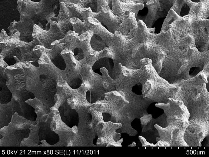
Sample prep on this sample was similar to the Pocillopora sample. Images were acquired on a Hitachi 4800 Field Emission Gun SEM. The skeleton has some interesting structure:


Here’s where a polyp would come from:

And the tip of one of those tines from the polyp area:

I zoomed in a bit on the crystal structure of the skeleton (from about the middle of the above pic):

Here’s another area of the skeleton that I zoomed in on. There are some very different structures here:

Check out these spherical features I found. …who’d of guessed?


0 Comments